
Page 129 - Registrar Orientation Manual 2016
P. 129
Reference:
1257
Effective date:
7 July 2015
Expiry date:
6 July 2018
Page:
12 of 35
Title:
Imaging Guidelines
Type:
Clinical Guideline
Version:
02
Authorising initials:
Pyelonephritis (Suspected)
In uncomplicated acute pyelonephritis imaging is not usually indicated.
There are several indications for imaging in suspected acute pyelonephritis:
• when the diagnosis is uncertain/equivocal
• if obstruction of the collecting system is suspected
• patients with suspected or known underlying anatomical predisposition
• recurrent acute pyelonephritis
• in ‘high risk’ patients who are more susceptible to occult infection or complications: this includes individuals with diabetes mellitus, elderly patients, patients who are immunosuppressed or on immunosuppressant therapy, renal transplant patients
• in patients who fail to respond to culture sensitive intravenous antibiotics within 72hrs Ultrasound is usually the appropriate first line investigation in such cases.
CT is more sensitive than ultrasound in diagnosing pyelonephritis or its complications and/or excluding other causes and may be used selectively in patients with no evidence of obstruction on ultrasound, if they are not improving (especially for ‘high risk’ patients).
Abdominal Masses and Organomegaly
The option of ultrasound should be considered (as it is a non-contrast investigation) but in patients with suspected malignancy abdominal CT is usually preferred over ultrasound unless the patient has renal impairment, a contraindication to contrast or suspected biliary obstruction.
Clinical urgency: Dependent on the acuity. Diagnostic imaging takes precedence over staging.
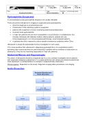
Aortic Dissection
Suspected Spontaneous Aortic Dissection
CXR
CT or TOE
(If patient is critically ill)
Normal
Type A
Type B
TOE/Angiography for coronary/functional assessment
Treat
Treat
Consider other diagnosis
