
Page 128 - Registrar Orientation Manual 2016
P. 128
Reference:
1257
Effective date:
7 July 2015
Expiry date:
6 July 2018
Page:
11 of 35
Title:
Imaging Guidelines
Type:
Clinical Guideline
Version:
02
Authorising initials:
Pancreatitis (Suspected)
The role of imaging in acute pancreatitis is to:
• exclude an underlying cause (e.g. gallstones)
• assess severity
• detect complications
• guide treatment of complications (e.g. fluid collection drainage)
Routine CT scan is not indicated. CT indications include:
• where diagnosis is in doubt
• clinically severe cases to assess degree of pancreatic necrosis
• failure to improve or sudden deterioration
• imaging complications of pancreatitis
Ultrasound may help determine the aetiology by assessing for gallstones and dilated ducts ERCP indications are shown in the pathway.
*MRCP and/or endoscopic US may help in determining the aetiology in difficult cases but may not be appropriate if an interventional ERCP is indicated anyway.
*MRCP or EUS
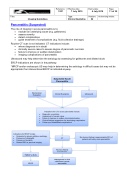
Suspected Acute Pancreatitis
Biochemical Evaluation
Clinical Evaluation
Ultrasound
Indications for CT in acute pancreatitis include:
• Diagnostic uncertainty.
• Assessment of severe cases.
• Failure to improve or sudden clinical deterioration.
• Clinical findings suggestive of a developing complication.
• Follow-up of established complications.
Indications for ERCP include:
• Suspected or proven gallstone aetiology.
• Presence of cholangitis.
• Presence of jaundice.
• Dilated CBD on previous imaging.
CT
No obvious finding in approximately 30% of patients with early acute pancreatitis.
Revised Atlanta Classification
Management of fluid collections in acute pancreatitis
ERCP
