
Page 124 - Registrar Orientation Manual 2016
P. 124
Reference:
1257
Effective date:
7 July 2015
Expiry date:
6 July 2018
Page:
7 of 35
Title:
Imaging Guidelines
Type:
Clinical Guideline
Version:
02
Authorising initials:
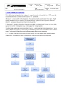
Cholecystitis (Suspected)
Plain abdominal radiography has no place in suspected acute cholecystitis but a CXR may help exclude a pulmonary condition as the cause of the symptoms.
Ultrasound is very specific in the diagnosis of acute cholecystitis, particularly if the signs of gall bladder wall thickening or oedema, peri-cholecystic fluid, gallstones and a positive ultrasonic Murphy's sign are all present but it has limited sensitivity.
If ultrasound is negative alternative diagnoses should be considered and if these are more likely appropriate further investigation (e.g. endoscopy) may be appropriate.
The Australian guidelines recommend that if there is a continuing high clinical suspicion of acute cholecystitis but a negative (or technically unsatisfactory) ultrasound a Tc-HIDA nuclear medicine scan is performed but we have more limited access to radio-isotope scanning.
A CT may help when the clinical picture is non-specific as it can detect other intra-abdominal inflammatory processes and when complications of acute cholecystitis are suspected.
Suspected Acute Cholecystitis
CXR
Abdominal USS
Positive for acute cholecystitis
Negative, but high clinical suspicion of acute cholecystitis, or equivocal/ technically inadequate US
Negative, low clinical suspicion
CT
Positive for acute cholecystitis
Negative
Consider alternative diagnoses
Treat
Peptic ulcer disease
Other non- traumatic acute abdominal pain
Endoscopy
Treat
